
Ultrasound for Dogs with Cancer: How It Works, What to Expect, and Why It's Recommended
Jun 25, 2026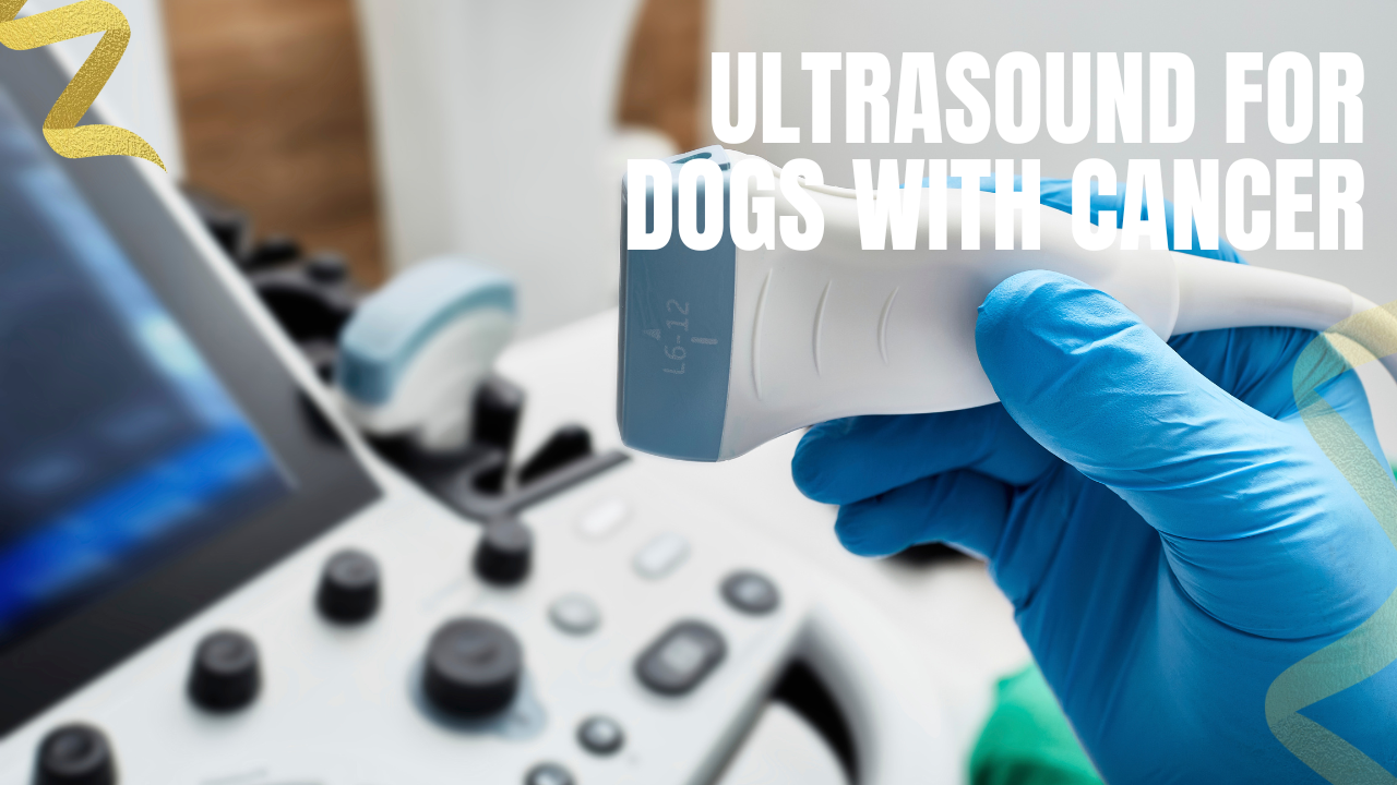
If your veterinarian has recommended an ultrasound for your dog, it can feel like one more worrying step in an already stressful process. But an ultrasound is one of the most useful, lowest-risk tools in all of veterinary medicine. It's painless, it doesn't use radiation, and in the world of cancer it does two big jobs: it helps find and characterize tumors, and it helps map out how far a known cancer has spread. Here's a clear guide to what an ultrasound is, why it matters so much in cancer care, and what to expect on the day.
What an Ultrasound Actually Is
An ultrasound (also called sonography) works by sending harmless, high-frequency sound waves into the body with a small handheld probe. Those waves bounce off the organs and tissues and return as echoes, which the machine converts into real-time, moving images. Think of it as a way for your veterinarian to look inside your dog's body, watching organs as they actually are, without a single incision.
A key point that often surprises owners: an ultrasound is not the same as an X-ray, and one doesn't replace the other. X-rays (radiographs) are best for dense structures like bones and the lungs, and they capture a single still snapshot. Ultrasound shines at the soft tissues, the detailed internal architecture of organs like the liver, spleen, and kidneys, and shows them moving in real time. The two are complementary, which is exactly why a cancer workup often includes both: chest X-rays to check the lungs, and an abdominal ultrasound to examine the belly.
What an Ultrasound Looks At
Most cancer-related ultrasounds are abdominal ultrasounds, and they give a thorough tour of the belly rather than focusing on just one spot. In a single scan, your veterinarian can evaluate the liver, gallbladder, spleen, kidneys, adrenal glands, pancreas, stomach and intestines, urinary bladder, lymph nodes, and reproductive organs such as the prostate. Scanning everything, even when there's a specific area of concern, helps ensure nothing is missed.
Ultrasound isn't limited to the abdomen, either. An echocardiogram is simply an ultrasound of the heart, used to investigate cardiac masses, and ultrasound can also help assess masses in the chest and certain structures in the neck.
Job One: Helping Find and Characterize Tumors
When a dog has vague signs of illness, abnormal bloodwork, or a mass your vet can feel during an exam, ultrasound is often the next step. It can locate a mass, pinpoint which organ it's growing from, measure it, and describe its texture and blood supply. For a dog facing possible surgery, this information is invaluable: it can clarify whether an operation is realistic, or sometimes spare a dog from surgery that wouldn't help.
There's an important honesty point here, though. An ultrasound cannot, on its own, diagnose cancer. It can reveal that something looks abnormal, but the way a mass appears on the screen does not reliably tell benign from malignant. A landmark study of splenic lesions found that the ultrasound appearance alone correlated poorly with the actual diagnosis, underscoring that imaging needs to be paired with an actual tissue sample to know what you're dealing with (Ballegeer et al., 2007). In other words, ultrasound tells you where and how big; cytology or biopsy tells you what.
The Standout Feature: Sampling Without Surgery
This is where ultrasound becomes genuinely powerful. Because it shows organs in real time, a veterinarian can watch a needle on the screen and guide it precisely into a mass or organ to collect cells — a procedure called an ultrasound-guided fine-needle aspirate (FNA). Sometimes a slightly larger needle is used to take a small core of tissue (a needle biopsy). Either way, your dog can have internal organs sampled without major surgery, which once would have required opening the abdomen.
These samples are then examined under a microscope by a pathologist, and they're frequently the key to an actual diagnosis. It's worth knowing the limits, though, because your veterinary team will be thinking about them too: a fine-needle sample collects only a small number of cells, and cytology doesn't always match what a full tissue biopsy would show.
In one study of liver aspirates, for example, the cytology results agreed with histopathology only some of the time (Wang et al., 2004). The practical upshot is that an FNA is an excellent, low-risk first step, but if the answer is unclear or doesn't fit the bigger picture, your vet may recommend a larger biopsy to confirm.
Job Two: Staging a Known Cancer
Once a cancer is diagnosed, the next question is how far it has gone — a process called staging. Staging guides treatment decisions and helps your veterinary team give you a realistic sense of prognosis, and abdominal ultrasound is a cornerstone of it (Vail et al., 2020). For lymphoma, for instance, a complete staging workup typically combines bloodwork, chest X-rays, an abdominal ultrasound to examine the spleen, liver, and abdominal lymph nodes, and often a bone marrow sample (North Carolina State University Veterinary Hospital, n.d.).
Here's a nuance that explains a recommendation owners sometimes find puzzling. For certain high-risk cancers, such as aggressive mast cell tumors, oncologists may recommend aspirating the spleen and liver even when those organs look completely normal on ultrasound. That's because microscopic spread can be present before it ever changes an organ's appearance — the same lesson from the splenic study above. Sampling normal-looking organs can catch early spread that imaging alone would miss, which can meaningfully change the treatment plan. So if your vet suggests sampling an organ that "looked fine," it's not overkill; it's thoroughness.
How Ultrasound Compares to CT and MRI
Ultrasound is fast, widely available, sedation-free for many dogs, and free of radiation, which makes it the natural first-line imaging tool for the abdomen. It does have limits: it can't see well through gas or bone, it's not the best way to evaluate the lungs, and image quality depends a great deal on the skill of the person performing it.
For some situations — mapping the full extent of a tumor before surgery or radiation, or detecting spread that ultrasound can miss — a CT or MRI scan may be more sensitive. These are more involved (they usually require general anesthesia) and aren't always necessary, but your oncologist may recommend one when the added detail would change the plan. None of this diminishes ultrasound's value; it simply has a role, and a very important one.
What to Expect on the Day
The good news for owners is that an ultrasound is one of the easiest procedures your dog will go through.
- Fasting. For an abdominal ultrasound, your dog will usually need an empty stomach, so expect instructions to withhold food for roughly 8 to 12 hours beforehand (water is generally fine). Food and gas in the digestive tract can block the view of nearby organs. If the bladder is a focus, your vet may ask you to prevent urination beforehand so it stays full.
- Shaving. A patch of fur on the belly is clipped. Hair traps air and blocks the sound waves, so bare skin (plus warm gel) gives a much clearer picture and better answers.
- Positioning and the scan itself. Your dog lies on their back or side in a padded trough while the veterinarian moves the probe across the belly. It's painless, and a full abdominal scan typically takes around 30 to 60 minutes.
- Sedation. Many dogs need none at all. Sedation may be offered for anxious or uncomfortable dogs, and it's generally recommended if an ultrasound-guided biopsy is being taken, to keep your dog still and safe.
- Afterward. Because the scan is non-invasive, there's little aftercare beyond a shaved patch and, if sedation was used, a little extra sleepiness. Your vet may also use repeat ("recheck") ultrasounds during treatment to see whether a tumor is shrinking and whether therapy is working.
The Bottom Line
An ultrasound won't, by itself, tell you whether your dog has cancer — that takes a tissue sample and a pathologist. But it does something almost nothing else can: it lets your veterinary team see inside your dog safely, find and measure masses, guide a needle to collect a diagnosis without surgery, and map how far a cancer has spread so the treatment plan fits the situation.
It's painless, radiation-free, and usually quick, which is a lot of value for very little burden on your dog. If your veterinarian has recommended one, it's almost always a smart, low-risk step toward getting the clearest possible answers — and the best possible plan.
This article is for educational purposes and reflects the current state of veterinary practice; it isn't a substitute for individualized veterinary advice. Always consult your veterinarian or a veterinary oncologist about the right diagnostic plan for your dog.
References
Ballegeer, E. A., Forrest, L. J., Dickinson, R. M., Schutten, M. M., Delaney, F. A., & Young, K. M. (2007). Correlation of ultrasonographic appearance of lesions and cytologic and histologic diagnoses in splenic aspirates from dogs and cats: 32 cases (2002–2005). Journal of the American Veterinary Medical Association, 230(5), 690–696. https://doi.org/10.2460/javma.230.5.690
Koehler, R. (2024, May 30). Dog ultrasounds: What are they, and why would a dog need one? PetMD. https://www.petmd.com/dog/procedure/dog-ultrasounds
North Carolina State University Veterinary Hospital. (n.d.). Cancer staging. Retrieved June 26, 2026, from https://hospital.cvm.ncsu.edu/services/small-animals/cancer-oncology/oncology/cancer-staging/
Vail, D. M., Thamm, D. H., & Liptak, J. M. (Eds.). (2020). Withrow and MacEwen's small animal clinical oncology (6th ed.). Saunders/Elsevier.
Wang, K. Y., Panciera, D. L., Al-Rukibat, R. K., & Radi, Z. A. (2004). Accuracy of ultrasound-guided fine-needle aspiration of the liver and cytologic findings in dogs and cats: 97 cases (1990–2000). Journal of the American Veterinary Medical Association, 224(1), 75–78. https://doi.org/10.2460/javma.2004.224.75
Reviewed by: Amber L. Drake, PhD
Dr. Amber L. Drake is a board-certified holistic health practitioner, canine clinical herbalist, educator, and founder of the Drake Dog Cancer Foundation and Drake Dog Academy. She is dedicated to helping pet parents better understand canine cancer, treatment options, nutrition, quality of life, and supportive care through compassionate, evidence-informed education. Her work combines professional training, practical resources, and firsthand insight from supporting thousands of dog families through the challenges of a cancer diagnosis.
Stay connected with news and updates!
Join our mailing list to receive the latest news and updates from our team.
Don't worry, your information will not be shared.
We hate SPAM. We will never sell your information, for any reason.