Could The Lump Or Abnormal Test Result Be Something Other Than Cancer?
Jul 25, 2026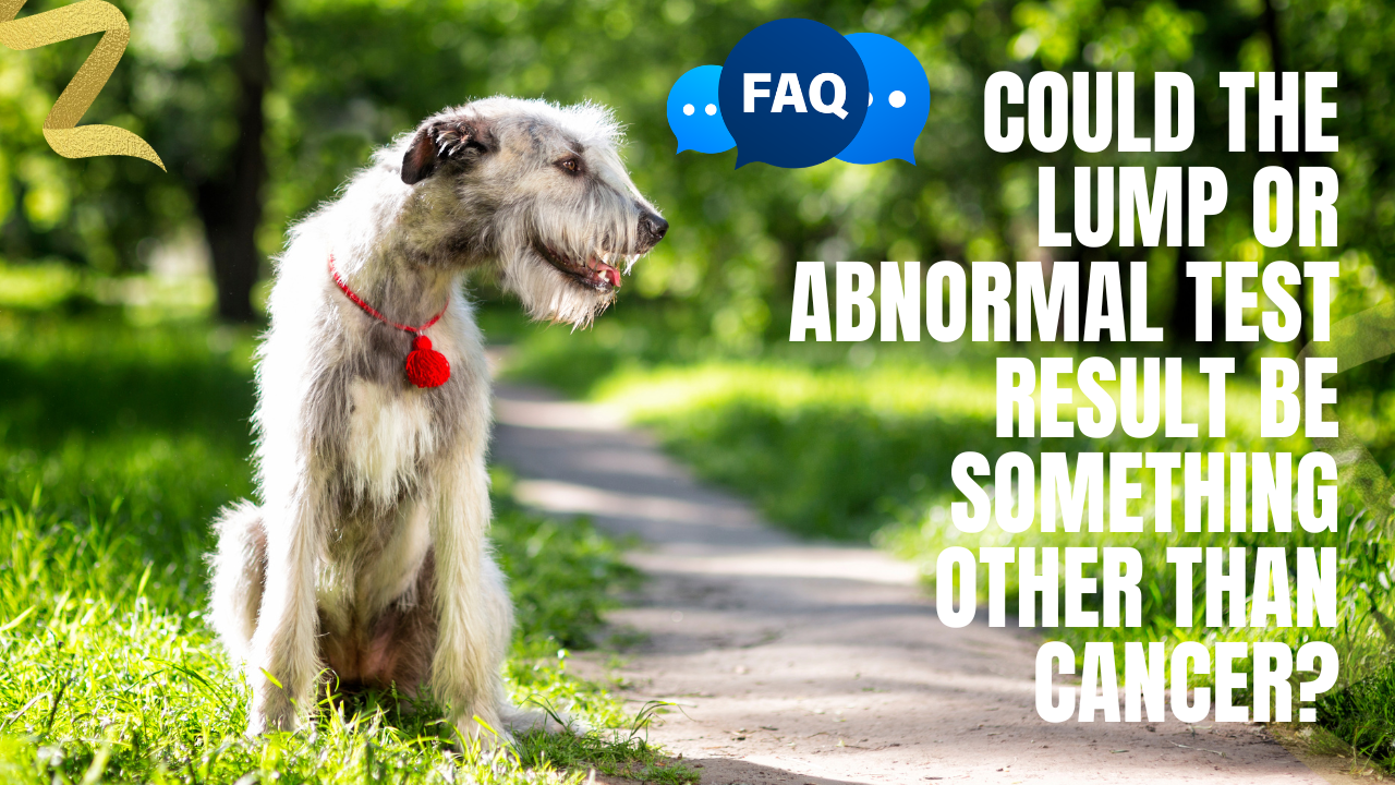
A lump, enlarged lymph node, abnormal scan, or unusual laboratory result can have many causes other than cancer.
Some findings are benign tumors, while others are caused by inflammation, infection, injury, fluid accumulation, hormonal disease, or normal age-related changes. However, it is usually impossible to determine what a lump is by appearance or touch alone. Even experienced veterinarians often need to examine cells or tissue under a microscope before they can provide a reliable diagnosis.
What Else Could A Lump Be?
A lump may be caused by several different conditions.
A Benign Tumor
A benign tumor is an abnormal growth that is not cancerous. Benign tumors do not typically spread to distant organs, although they can still enlarge, become irritated, interfere with movement, or press on nearby structures.
Common examples include:
-
Lipomas
-
Sebaceous gland adenomas
-
Histiocytomas
-
Papillomas
-
Fibromas
-
Skin tags
-
Some mammary tumors
-
Certain hair-follicle tumors
Lipomas are among the most common benign tumors in dogs. They are made of fatty tissue and are often soft, rounded, slow-growing, and movable beneath the skin. However, these characteristics are not enough to confirm that a lump is a lipoma.
A Cyst
A cyst is a pocket within the tissue that contains fluid, keratin, oil, or another material.
Cysts may:
-
Feel firm or soft
-
Develop a small opening
-
Release thick or oily material
-
Become inflamed
-
Rupture
-
Become infected
-
Return after being drained
Although many cysts are benign, a veterinarian should evaluate them because some tumors can resemble cysts externally.
An Abscess Or Infection
An abscess is a pocket of infection that may form after:
-
A bite
-
A puncture wound
-
A foreign object
-
A skin infection
-
An infected anal gland
-
A dental infection
-
A surgical complication
An abscess may be warm, painful, red, swollen, or filled with pus. The dog may also develop fever, lethargy, poor appetite, or discomfort.
Infections can also enlarge lymph nodes or create abnormalities in internal organs that resemble cancer on imaging.
Inflammation
Inflammation can cause swelling, nodules, thickened tissue, or abnormal findings on diagnostic tests.
Possible causes include:
-
Allergic reactions
-
Insect bites or stings
-
Autoimmune disease
-
Pancreatitis
-
Inflammatory bowel disease
-
Granulomas
-
Injection-site reactions
-
Chronic irritation
-
Foreign-body reactions
Inflammatory cells may also dominate a fine-needle aspiration sample and make it difficult to determine whether an underlying tumor is present.
A Hematoma Or Seroma
A hematoma is a collection of blood beneath the skin or within tissue. It may develop after trauma, surgery, a bite, vigorous activity, or a blood-clotting problem.
A seroma is a pocket of clear fluid that can form after surgery or tissue injury.
These swellings may resemble tumors, particularly when the injury was not witnessed.
Scar Tissue Or A Reaction To Injury
Scar tissue, calluses, pressure sores, and healing injuries can sometimes feel like firm masses.
Dogs may develop thickened areas over:
-
Elbows
-
Hocks
-
The chest
-
Surgical sites
-
Areas of repeated pressure or friction
However, a lump near an old injury or incision should not automatically be assumed to be scar tissue.
An Enlarged Lymph Node
Lymph nodes may enlarge because of:
-
Infection
-
Inflammation
-
Immune stimulation
-
Dental disease
-
Skin disease
-
Tick-borne illness
-
Lymphoma
-
Metastatic cancer
An enlarged lymph node does not automatically mean cancer. Conversely, a lymph node can contain cancer cells without being noticeably enlarged.
Sampling the node is often necessary to determine why it has changed.
A Hernia
A hernia occurs when tissue or an organ pushes through an opening in the surrounding muscle or connective tissue.
Depending on the location, a hernia may appear as a soft or firm swelling near the:
-
Belly button
-
Groin
-
Diaphragm
-
Perineal area
-
Previous surgical site
Some hernias are stable, while others require urgent treatment if tissue becomes trapped or its blood supply is compromised.
A Normal Anatomical Structure
Pet parents sometimes discover a normal structure that feels unfamiliar, such as:
-
A nipple
-
A rib
-
The end of the sternum
-
A salivary gland
-
A lymph node
-
A bony prominence
-
Normal fatty tissue
A veterinary examination can help distinguish normal anatomy from a new mass.
Can A Cancerous Lump Look Benign?
Yes. Some malignant tumors can be soft, movable, slow-growing, painless, or present for months without causing noticeable illness.
For example, soft tissue sarcomas often appear as soft or firm lumps beneath the skin. Many grow slowly and are not painful, even though they can invade surrounding tissue microscopically.
A mast cell tumor may also change size, become swollen and then shrink, or resemble an insect bite, cyst, fatty lump, or harmless skin growth.
This is why appearance and texture cannot reliably separate benign tumors from cancer.
Can A Benign Lump Look Concerning?
Yes. Benign growths can:
-
Grow rapidly
-
Bleed
-
Become ulcerated
-
Feel firm
-
Become inflamed
-
Interfere with walking
-
Develop in a difficult location
-
Look irregular
-
Return after removal
Inflammation, infection, and trauma can also make a harmless mass look more concerning.
Testing is needed to determine the cause rather than relying only on how dramatic the lump appears.
Can A Veterinarian Tell By Feeling The Lump?
A veterinarian may develop a list of likely possibilities based on the lump’s:
-
Location
-
Size
-
Shape
-
Texture
-
Mobility
-
Growth pattern
-
Surface appearance
-
Relationship to nearby tissues
However, examination alone usually cannot provide a definitive diagnosis.
Cornell notes that although some masses have characteristic appearances or locations, there is no way to know with complete certainty what a mass is simply by looking at or feeling it.
What Is A Fine-Needle Aspiration?
A fine-needle aspiration, commonly called an FNA, uses a small needle to collect cells from a lump, lymph node, organ, or fluid-filled area.
The cells are placed on microscope slides and examined through cytology.
An FNA can sometimes identify:
-
Lipomas
-
Mast cell tumors
-
Lymphoma
-
Inflammation
-
Infection
-
Cysts
-
Certain round-cell tumors
-
Some metastatic cancers
Fine-needle aspiration is one of the most frequently used methods for evaluating lumps and bumps. It is generally quick and minimally invasive, and many superficial masses can be sampled without sedation.
Can A Fine-Needle Aspiration Rule Out Cancer?
Not always.
An FNA may be inconclusive when:
-
Too few cells are collected
-
The sample contains mostly blood
-
The needle enters an area of inflammation or tissue death
-
The tumor does not release cells easily
-
The wrong part of the mass is sampled
-
The cells look similar to several possible diseases
-
Tumor grade cannot be determined from individual cells
A nondiagnostic or benign-appearing aspiration does not always completely rule out cancer.
Cornell advises that cytology should not be overinterpreted and that major treatment decisions should rely on cytology only when the sample provides a sufficiently definitive diagnosis.
When Is A Biopsy Needed?
A biopsy removes a larger piece of tissue or the entire mass so a veterinary pathologist can examine the cells and the way they are arranged.
A biopsy may be recommended when:
-
The FNA is inconclusive
-
The lump continues to grow
-
The clinical appearance does not match the cytology result
-
The tumor type cannot be identified
-
Tumor grade is needed
-
Major surgery is being considered
-
The mass is located in a complicated area
-
The veterinarian needs to plan surgical margins
-
A previous sample did not provide enough information
Histopathology can often provide a more complete classification because it evaluates tissue architecture in addition to individual cells.
Should Every Lump Be Removed?
Not necessarily.
Some lumps may be safely monitored after a reliable diagnosis has been obtained. Others should be removed because they are:
-
Cancerous
-
Rapidly growing
-
Painful
-
Bleeding
-
Infected
-
Repeatedly irritated
-
Interfering with movement
-
Located where future growth would make surgery difficult
-
Diagnostically uncertain
The decision should be based on the diagnosis, location, growth pattern, surgical risks, and your dog’s overall health.
Avoid assuming that a lump is harmless and monitoring it indefinitely without first discussing sampling with your veterinarian. Delaying testing may allow a malignant mass to become larger and require more extensive surgery.
What Changes Make A Lump More Concerning?
Have a veterinarian evaluate any new or changing lump. Features that may justify more urgent assessment include:
-
Rapid growth
-
Sudden change in size
-
Firm attachment to deeper tissue
-
Irregular shape
-
Ulceration
-
Bleeding
-
Discharge
-
Redness or warmth
-
Pain
-
Repeated swelling and shrinking
-
Dark or changing pigmentation
-
Hair loss over the lump
-
Difficulty walking, eating, breathing, urinating, or defecating
-
Enlarged lymph nodes
-
Weight loss or reduced appetite
-
Multiple new masses
These signs do not prove that the lump is cancerous, but they increase the importance of obtaining a diagnosis.
Could An Abnormal Blood Test Mean Something Other Than Cancer?
Yes. Bloodwork abnormalities are rarely specific to cancer.
Changes may be caused by:
-
Infection
-
Inflammation
-
Dehydration
-
Kidney disease
-
Liver disease
-
Hormonal disorders
-
Blood loss
-
Immune-mediated disease
-
Medication effects
-
Nutritional deficiencies
-
Toxin exposure
-
Bone-marrow disease
-
Cancer
For example, anemia, high calcium, elevated liver values, or abnormal white blood cell counts may occur with cancer, but each can also have multiple noncancerous causes.
Bloodwork may provide clues, but it usually cannot confirm most solid tumors by itself.
Could An Abnormal X-Ray Or Ultrasound Be Something Other Than Cancer?
Yes. Imaging abnormalities can represent:
-
Cysts
-
Benign nodules
-
Abscesses
-
Inflammation
-
Scar tissue
-
Organ enlargement
-
Congenital changes
-
Fluid accumulation
-
Blood clots
-
Infection
-
Age-related changes
-
Cancer
Imaging can identify the location, size, and appearance of an abnormality, but cells or tissue may still be needed to determine exactly what it is.
A suspicious finding should not automatically be labeled cancer without appropriate confirmation whenever sampling is safe and clinically useful.
Could A Positive Cancer-Screening Test Be Wrong?
Cancer-screening tests may estimate the likelihood that cancer-related signals are present, but they do not always identify the tumor’s location or replace diagnostic testing.
Possible results include:
-
True positive
-
False positive
-
True negative
-
False negative
-
A result that requires further investigation
Newer blood- and urine-based liquid biopsy tests are being studied and may assist with detecting some cancers. However, current evidence does not support using them as a universal replacement for tissue biopsy, and early-stage detection remains an evolving area of veterinary medicine.
A positive screening result generally means further examination and diagnostic testing are needed. A negative result does not rule out every cancer.
What Should I Do If The Test Result Is Inconclusive?
Ask your veterinarian:
-
Was enough material collected?
-
Should the aspiration be repeated?
-
Should a different part of the lump be sampled?
-
Should the slides be reviewed by a clinical pathologist?
-
Is a tissue biopsy recommended?
-
Would imaging help guide the sample?
-
Is complete removal appropriate?
-
How soon should the lump be reassessed?
-
What changes should I monitor at home?
Do not interpret an inconclusive result as confirmation that the mass is harmless.
Should I Get A Second Pathology Opinion?
A second review may be helpful when:
-
The diagnosis is rare
-
The report is uncertain
-
The result does not match the mass’s behavior
-
Major or irreversible treatment is being considered
-
The oncologist recommends additional review
-
Different tests provide conflicting information
-
The sample is difficult to classify
Existing slides or tissue blocks can often be sent to another veterinary pathologist without repeating the procedure.
How Should I Monitor A Lump At Home?
Keep a record of:
-
Location
-
Length and width
-
Shape
-
Texture
-
Color
-
Mobility
-
Surface changes
-
Pain or sensitivity
-
Bleeding or discharge
-
Date first noticed
-
Rate of growth
Photograph the lump beside a ruler or measuring tape under similar lighting each time.
Home monitoring is useful, but it should not replace veterinary evaluation or diagnostic testing.
Questions To Ask Your Veterinarian
-
What are the most likely causes of this lump?
-
Could it be an infection, cyst, or benign tumor?
-
Should it be sampled now?
-
Is a fine-needle aspiration appropriate?
-
Should the sample be sent to a pathologist?
-
What would an inconclusive result mean?
-
Would a biopsy provide more information?
-
Could removing it now make surgery easier?
-
Is it safe to monitor?
-
How frequently should I measure it?
-
What changes should prompt an immediate recheck?
-
Could the abnormal bloodwork have a noncancerous cause?
-
Does the imaging finding need to be sampled?
-
Should we repeat the test before making a treatment decision?
The Most Important Takeaway
A lump or abnormal test result does not automatically mean your dog has cancer. Benign tumors, cysts, infections, inflammation, injury, and many other conditions can cause similar findings.
At the same time, a lump cannot be reliably declared harmless based only on its appearance, softness, movement, or slow growth.
The safest approach is to have new or changing lumps examined and, when appropriate, sampled through fine-needle aspiration or biopsy. Obtaining a diagnosis early can prevent unnecessary worry when a lump is benign and allow earlier treatment when it is not.
References
American Veterinary Medical Association. (2025). Cancer in pets. https://www.avma.org/resources/pet-owners/petcare/cancer-pets
Cornell University College of Veterinary Medicine. (2025). Lipomas in dogs. https://www.vet.cornell.edu/departments-centers-and-institutes/riney-canine-health-center/canine-health-information/lipomas-dogs
Cornell University College of Veterinary Medicine. (n.d.-a). Diagnosis and staging. Retrieved July 16, 2026, from https://www.vet.cornell.edu/departments-centers-and-institutes/sprecher-institute-comparative-cancer-research/treatment-strategies/diagnosis-and-staging
Cornell University College of Veterinary Medicine. (n.d.-b). Skin lumps and bumps. Retrieved July 16, 2026, from https://riney.vet.cornell.edu/member-benefits-health-tips/skin-lumps-bumps-month-november-1
Cornell University College of Veterinary Medicine. (n.d.-c). Soft tissue sarcomas in dogs. Retrieved July 16, 2026, from https://www.vet.cornell.edu/departments-centers-and-institutes/riney-canine-health-center/canine-health-information/soft-tissue-sarcomas-dogs
Flory, A., Kruglyak, K. M., Tynan, J. A., McLennan, L. M., Rafalko, J. M., Fiaux, P. C., Hernandez, G. E., Marass, F., Nakashe, P., Ruiz-Perez, C. A., Schillebeeckx, C., Smit-McBride, Z., Baltzley, S. J., Lau, J. W., Burnett, K. R., Chudoba, I., Conkling, P., Tannahill, A., Jordan, L. A., & Grosu, D. S. (2024). Clinical validation of a blood-based liquid biopsy test for cancer detection in dogs. Journal of the American Veterinary Medical Association, 262(5), 712–721. https://doi.org/10.2460/javma.23.10.0564
VCA Animal Hospitals. (2024). Adipose tumors: Lipomas in dogs. https://vcahospitals.com/know-your-pet/adipose-lipoma-tumors
VCA Animal Hospitals. (2026a). Cysts. https://vcahospitals.com/know-your-pet/cysts
VCA Animal Hospitals. (2026b). Cytology: General. https://vcahospitals.com/know-your-pet/cytology---general
VCA Animal Hospitals. (2026c). Cytology, biopsy, and histopathology. https://vcahospitals.com/know-your-pet/cytology-histopathology-biopsies
VCA Animal Hospitals. (n.d.). Fine needle aspiration. Retrieved July 16, 2026, from https://vcahospitals.com/know-your-pet/cytology-fine-needle-aspiration-fna
Reviewed by: Amber L. Drake, PhD
Dr. Amber L. Drake is a board-certified holistic health practitioner, canine clinical herbalist, educator, and founder of the Drake Dog Cancer Foundation and Drake Dog Academy. She is dedicated to helping pet parents better understand canine cancer, treatment options, nutrition, quality of life, and supportive care through compassionate, evidence-informed education. Her work combines professional training, practical resources, and firsthand insight from supporting thousands of dog families through the challenges of a cancer diagnosis.
Stay connected with news and updates!
Join our mailing list to receive the latest news and updates from our team.
Don't worry, your information will not be shared.
We hate SPAM. We will never sell your information, for any reason.