
Mast Cell Tumors
The Most Common Malignant Skin Cancer
Mast cell tumors are the most common malignant skin tumor in dogs, and the single most important thing to understand about them is that they are not one disease with one outlook. They range from a lump that surgery removes permanently to an aggressive cancer needing serious treatment.
Which one your dog has is determined by grade, and grade comes from a pathologist examining the removed tumor. Nothing about how the lump looks, how big it is, or how long it has been there reliably predicts it.
Here is the encouraging part: the majority of mast cell tumors in dogs are low-grade, and for a low-grade tumor removed completely, surgery frequently ends the matter for good. Many dogs treated for a mast cell tumor go on to live a full life and die years later of something entirely unrelated.
If you are waiting on a pathology report, that wait is genuinely the hard part, and the odds are in your favor.

What Mast Cell Tumors Are
Mast cells are normal immune cells found throughout the body, particularly in skin and connective tissue. They are packed with granules containing histamine and other active substances, and they release those granules during allergic reactions — which is why an insect sting swells.
A mast cell tumor is a mass of these cells. Because the cells still contain their granules, the tumor can release histamine into the surrounding tissue and bloodstream. That release, called degranulation, explains most of what makes this cancer behave strangely.
Degranulation is why a mast cell tumor may swell, redden, and become itchy when handled. It is why some tumors get noticeably bigger and smaller from week to week. And it is why a tumor on the skin can cause stomach problems, since circulating histamine drives acid production and can lead to ulcers.
Do not squeeze, poke, or repeatedly handle a suspected mast cell tumor. Manipulation triggers degranulation and makes the local swelling worse.
The Tumor That Looks Like Anything
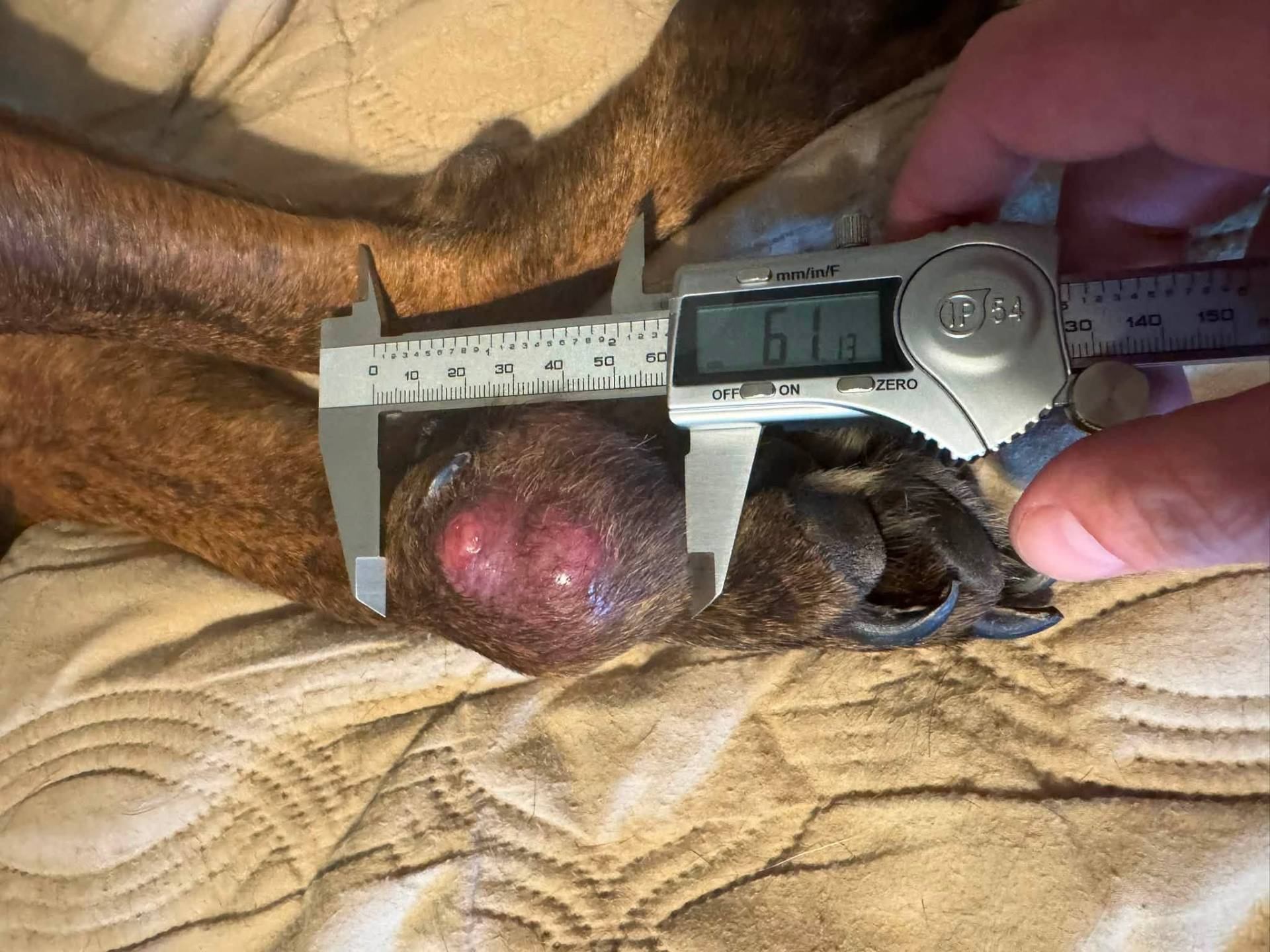
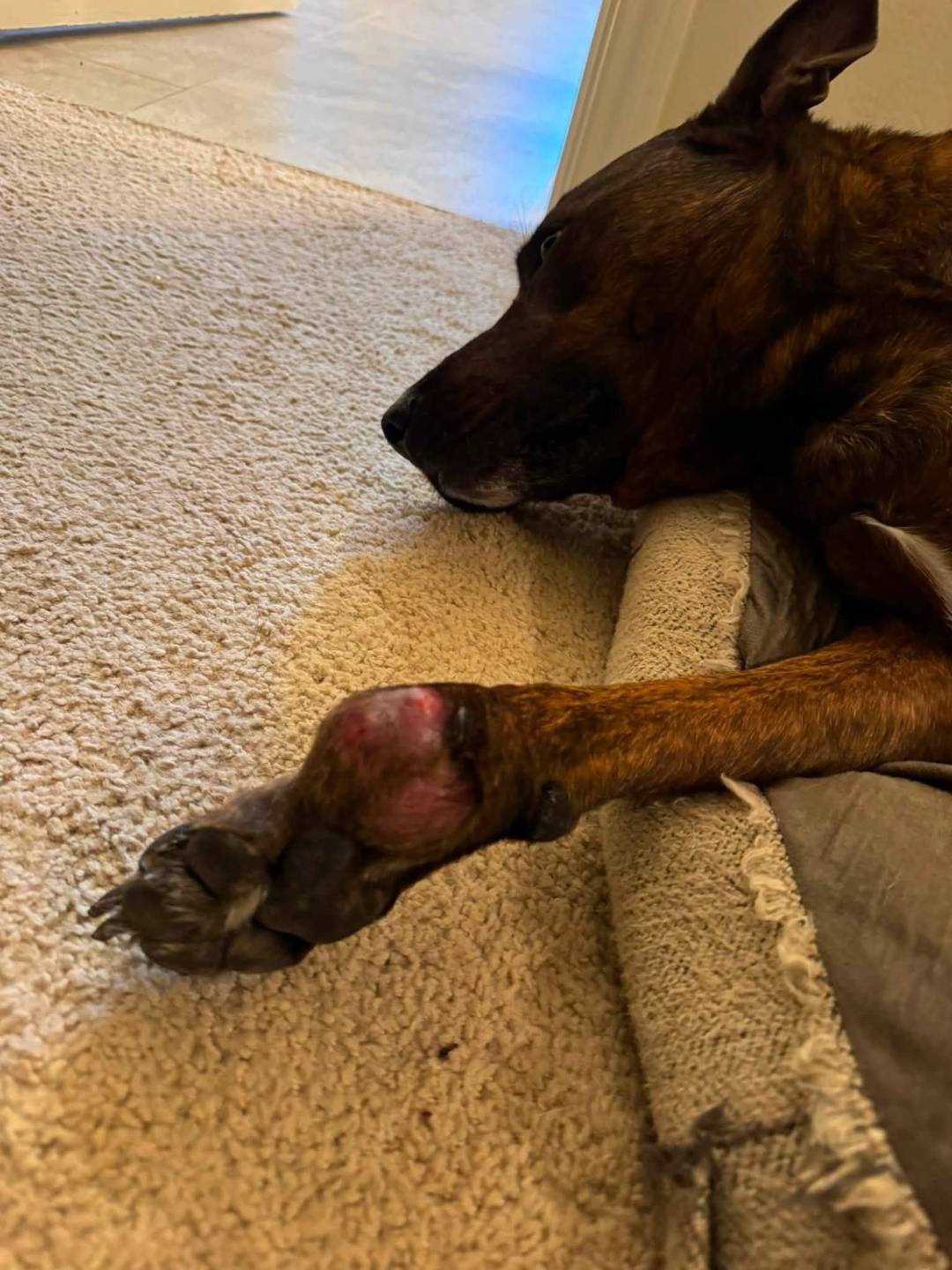
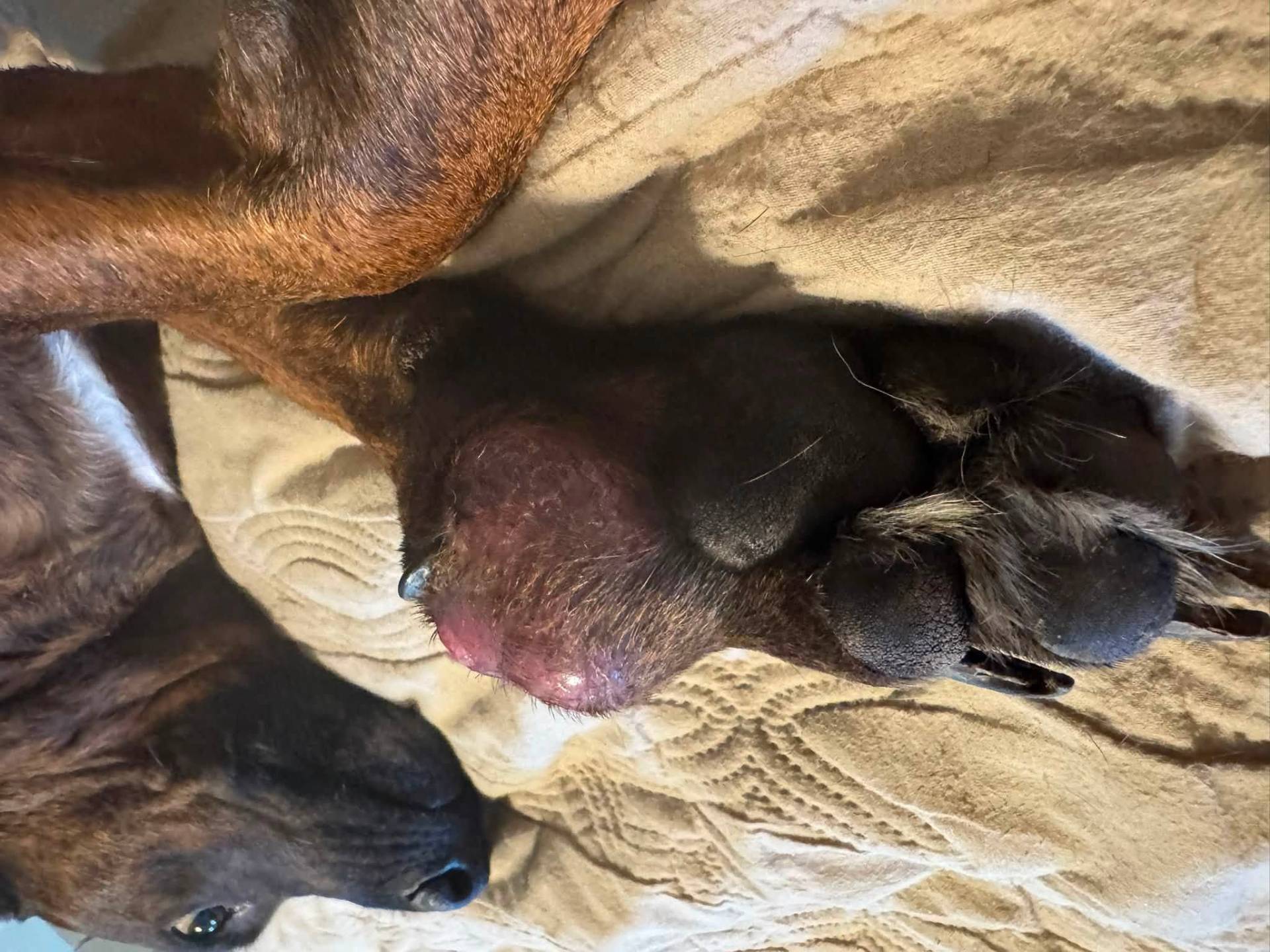
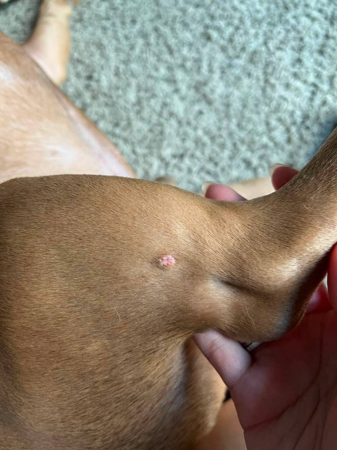
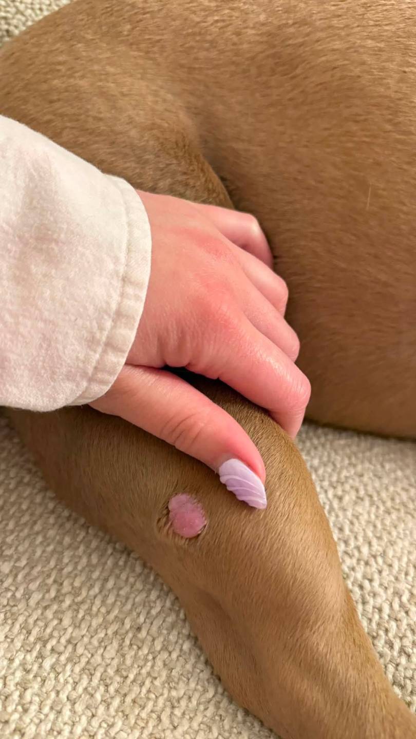
Mast cell tumors have no characteristic appearance. They can look like a wart, a fatty lump, an insect bite, a raised red sore, a soft squishy mass under the skin, or an area of thickened skin. They can be present for years without changing or appear and grow within weeks.
This is the practical consequence, and it is the most useful thing on this page:
Any new lump on your dog deserves a needle sample, not a watch-and-wait. Fine needle aspiration takes seconds, usually needs no sedation, and identifies mast cell tumors reliably because the granules are distinctive under a microscope. A lump that gets dismissed as a fatty tumor by appearance alone is the most common way this diagnosis gets delayed.
Aspirating lumps is cheap and quick. Assuming is not.
Read More: What Is a Fine Needle Aspirate?Images of Mast Cell Tumors from Our Pet Parents
Signs to Look For
Local signs:
- A lump anywhere on or under the skin, of any appearance
- A mass that changes size, sometimes swelling then subsiding
- Redness, swelling, or itching after the lump is touched
- Ulceration or bleeding of the surface
- Your dog licking or scratching persistently at one spot
Systemic signs, from histamine released into the bloodstream:
- Vomiting, sometimes with blood
- Black, tarry stool, indicating bleeding in the stomach or intestines
- Reduced appetite
- Lethargy
- Abdominal discomfort
Stomach ulceration is common enough with mast cell tumors that many oncologists treat for it preventively. Black tarry stool or vomiting blood warrants prompt veterinary attention.

Which Dogs Are Most Commonly Affected
Boxers, Boston Terriers, Bulldogs, Pugs, Labrador Retrievers, Golden Retrievers, Weimaraners, Rhodesian Ridgebacks, and Shar Peis are all diagnosed more often than average.
Breed influences behavior as well as risk. Boxers and Pugs develop mast cell tumors frequently but tend to develop low-grade ones. Shar Peis tend toward higher-grade tumors and at younger ages.
Most dogs are diagnosed around eight or nine years old, though they occur at any age.
How Mast Cell Tumors are Diagnosed
Fine needle aspiration usually provides the diagnosis. Mast cells have distinctive granules that are readily recognized on cytology, so a mast cell tumor is one of the few cancers frequently identified before any surgery happens. This is a real advantage, because it allows surgery to be planned properly the first time.
Histopathology is required for grading. Cytology tells you it is a mast cell tumor; only examination of the removed tissue tells you which kind. This is why the pathology report matters so much and why waiting for it is unavoidable.
Staging assesses whether the tumor has spread. Depending on the situation, this may include sampling the nearby lymph node, abdominal ultrasound with sampling of the spleen and liver, and bloodwork. Some centers use sentinel lymph node mapping to identify precisely which node drains the tumor.
How much staging is appropriate depends on risk. A small, low-grade tumor completely removed may not warrant an extensive workup, while a high-grade tumor does. Ask your veterinarian what they recommend for your dog specifically and why.
Understanding Your Pathology Report
This report determines your dog's treatment and outlook, and it arrives full of terminology nobody explains. Here is what to look for.
Grade
Two grading systems are in use, and most laboratories now report both.
The Patnaik system uses three grades:
- Grade I — well differentiated. Low risk of spread. Complete removal is usually the whole treatment.
- Grade II — intermediate. Historically the frustrating category, because it covered tumors that behaved very differently from one another.
- Grade III — poorly differentiated. High risk of spread, and aggressive.
The Kiupel system uses two tiers, and was developed specifically to resolve the ambiguity of Patnaik Grade II:
- Low grade — favorable, with median survival typically well over two years and many dogs living out a normal life span.
- High grade — aggressive, with median survival commonly reported at around four months without further treatment.
If your report gives both, the Kiupel result generally carries more weight, particularly where Patnaik says Grade II.
Mitotic count
This is the number of actively dividing cells the pathologist counted, and it is one of the strongest predictors on the whole report, sometimes more informative than grade.
A low count is favorable. A count above five is associated with significantly shorter survival and generally prompts more aggressive treatment. If your report lists a mitotic count, ask your veterinarian what it means in your dog's case.
Margins
This describes whether the tumor was fully removed, and it is reported as complete, incomplete, or narrow. It tells you the risk of the tumor regrowing at that site.
Complete margins on a low-grade tumor is the best possible outcome and usually means no further treatment. Incomplete margins mean tumor cells were left behind and something further is needed, a second surgery to remove more tissue, or radiation to the site.
Incomplete margins are common and are not a surgical failure. Mast cell tumors extend microscopically well beyond what can be seen or felt, and some locations simply do not have spare skin.
Additional testing
Some reports include further markers:
- Ki-67 measures how fast cells are proliferating and adds prognostic information.
- c-KIT mutation testing looks for a specific genetic change present in a minority of mast cell tumors. It matters practically: tumors carrying this mutation often respond well to a class of targeted drugs.
If your dog has a high-grade tumor or one that has recurred, ask whether c-KIT testing has been done, because it can open a treatment option.
Treatment
Surgery
Surgery is the primary treatment and, for most low-grade tumors, the only one needed.
Because mast cell tumors extend invisibly beyond their apparent edges, surgeons remove a margin of normal-looking tissue around and beneath the tumor. The traditional standard was three centimeters in every direction plus a layer of tissue underneath; current evidence supports somewhat narrower margins for low-grade tumors. The result is an incision considerably larger than the lump, which surprises many families.
Getting the first surgery right matters more than in almost any other canine cancer. A tumor removed with clean margins is often finished. A tumor removed narrowly requires a second operation into scarred tissue, which is harder and removes more. If a mast cell tumor is diagnosed by aspiration beforehand, ask whether your dog would benefit from a surgeon experienced with them.
Radiation
Radiation is highly effective for tumor cells left behind after incomplete removal, and it achieves excellent long-term control for low and intermediate grade tumors in that situation. It is also used where surgery is not possible because of location.
Medical treatment
Chemotherapy is recommended for high-grade tumors, tumors that have spread, and tumors with concerning features such as a high mitotic count. Vinblastine with prednisone and lomustine are commonly used.
Targeted drugs — toceranib and masitinib — block signals that drive mast cell tumor growth. Toceranib was the first drug approved specifically for cancer in dogs. These work particularly well in tumors carrying the c-KIT mutation and are given as tablets at home.
Prednisone has direct activity against mast cell tumors as well as reducing inflammation.
Injectable treatment into the tumor is an approved option for certain non-spread tumors, particularly in locations where surgery would be difficult. It destroys the tumor in place and leaves a wound that requires attention while it heals. Ask whether it suits your dog's situation.
Antihistamines and stomach protection
Nearly every dog with a mast cell tumor is prescribed antihistamines and a stomach-protecting medication. This is not incidental; it counteracts the histamine the tumor releases, protects against ulcers, and reduces the risk of complications during surgery. Take it as directed, including in the period before an operation.
Integrative and supportive care
Standard supportive care matters here: keeping your dog eating, managing any stomach upset, and protecting a surgical site while it heals.
Be careful with supplements. Anything affecting clotting is relevant around surgery, and some products marketed for allergies or immune support have unclear interactions with the antihistamine and steroid medications your dog is likely taking. Tell your veterinary team everything your dog receives.
No supplement, herb, or diet has been shown to treat mast cell tumors in dogs.
Comfort care alone
For a dog with widespread high-grade disease, or an older dog with other serious conditions, comfort-focused care is a legitimate path. It generally involves steroids, antihistamines, stomach protection, and pain relief, which together can keep a dog comfortable for a meaningful period. Discuss this openly with your veterinarian if aggressive treatment does not feel right for your dog.
What Treatment is Actually Like for Your Dog
Surgery is the most common experience, and for most dogs it is straightforward. Expect an incision considerably larger than the lump was, ten to fourteen days of restricted activity, and a recovery collar or surgical suit to prevent licking. Most dogs are comfortable within days on pain medication.
Radiation, when needed, involves a series of brief sessions under light anaesthesia. Skin in the treated area typically becomes red and irritated, similar to sunburn, and heals over several weeks.
Chemotherapy for dogs is dosed for quality of life rather than for eliminating the disease at any cost. Most dogs experience no significant side effects, and those who do usually have mild nausea or reduced appetite for a day or two after treatment. Most do not lose their coat. Our page on what chemotherapy is really like for dogs covers this in more depth.
Targeted tablets are given at home, with regular bloodwork to monitor. Side effects can include digestive upset, and dose adjustments are common.
Understanding Prognosis
Outlook depends almost entirely on grade, and the two ends of the range are very far apart.
Low-grade tumors removed completely carry an excellent prognosis. Median survival exceeds two years, and in practice many of these dogs are permanently free of the disease and live a normal life span. For this group — which is most dogs — a mast cell tumor is a problem that gets solved.
High-grade tumors are serious. Median survival is commonly reported at around four months without further treatment, and chemotherapy is recommended to extend that. Some dogs respond well and considerably exceed it.
Other factors that shift the picture: a mitotic count above five, incomplete margins, spread to lymph nodes or organs, recurrence after previous surgery, and tumors in certain locations such as the nail bed or internal organs.
If your dog's report says low grade with complete margins and a low mitotic count, that is genuinely good news and it is reasonable to feel relieved.
What It Costs
Costs vary by region and practice type. Ask for a written estimate.
- Aspiration and cytology: approximately $150 to $400
- Surgical removal: approximately $800 to $3,000, depending on size and location
- Histopathology: approximately $150 to $400
- Staging: approximately $500 to $1,500
- Radiation: approximately $4,000 to $8,000
- Chemotherapy: approximately $2,000 to $5,000
- Targeted tablets: several hundred dollars per month, ongoing
Compared with most cancers on this site, the common scenario here, aspirate, remove, send for histopathology, is relatively affordable.
Living with It Day to Day
Check your dog regularly for new lumps. Dogs who develop one mast cell tumor are more likely to develop others, and a meaningful proportion have more than one at diagnosis.
This is less alarming than it sounds. Multiple mast cell tumors are usually independent new tumors rather than the original one spreading, and dogs, particularly Boxers and Pugs, can have several removed over the years and do perfectly well. Each new lump is aspirated and assessed on its own.
Run your hands over your dog once a month. Some families keep a simple body map noting the location and size of anything found, which makes it easy to tell a new lump from one already checked.
Report to your veterinary team: any new lump, changes in an existing one, vomiting or black tarry stool, reduced appetite, or swelling near a previous surgical site.
Questions to Ask Your Veterinarian
- What grade is my dog's tumor under each system used?
- What was the mitotic count, and what does it mean here?
- Were the margins complete? If not, what are the options?
- How much staging does my dog need, and why that much?
- Is further treatment recommended, or is surgery sufficient?
- Should c-KIT testing be run?
- Should my dog be on antihistamines and stomach protection long term?
- How often should we recheck, and what should I watch for at home?
Thinking About Quality of Life
For most dogs, this section will not apply; a low-grade tumor removed completely is a finished problem, and the appropriate response is relief rather than planning.
For dogs with high-grade or widespread disease, quality of life becomes the guiding measure. Watch for appetite, comfort, nausea, energy, and engagement with the family. Stomach ulceration is a particular source of hidden discomfort in this cancer, so reduced appetite or black stool deserves attention rather than acceptance.
Scoring your dog weekly across a few consistent dimensions gives you a record more reliable than memory. If treatment stops working, ask your veterinary team what to expect and what the plan should be, ideally before you need it.
Common Questions
Is a mast cell tumor always serious? No. Most are low-grade, and complete surgical removal of a low-grade mast cell tumor frequently ends the problem permanently. A minority are high-grade and require aggressive treatment. Grade is what distinguishes them, and only histopathology determines it.
Why is the incision so much bigger than the lump was? Because mast cell tumors extend microscopically beyond their visible edges. Surgeons remove a margin of normal-looking tissue around and beneath the tumor to capture those cells. A generous first surgery is what makes a second one unnecessary.
The margins were incomplete. What now? This is common and manageable. Options are a second surgery to remove more tissue or radiation to the site, and radiation achieves excellent control for low and intermediate grade tumors in this situation. Discuss which suits your dog's tumor location and grade.
My dog has several lumps. Does that mean it has spread? Usually not. Dogs prone to mast cell tumors often develop multiple independent ones, which is different from a single tumor spreading. Each is aspirated and assessed separately, and many dogs have several removed over the years without their outlook worsening.
Why is my dog on antihistamines for cancer? Because mast cell tumors release histamine, which causes stomach ulcers and can trigger reactions during surgery. Antihistamines and stomach-protecting medication counteract this and are standard alongside treatment.
Can a mast cell tumor be removed for good? Often, yes. A low-grade tumor removed with complete margins frequently never returns, and those dogs commonly live out a normal life span. High-grade tumors are more likely to recur or spread and generally need treatment beyond surgery.
The lump changes size. Is that meaningful? It can be characteristic. Mast cell tumors sometimes swell and subside as they release histamine, and a mass that waxes and wanes is a recognized feature. Have any changing lump aspirated rather than watching it.
The Research
Blackwood L, Murphy S, Buracco P, et al. European consensus document on mast cell tumours in dogs and cats. Vet Comp Oncol. 2012;10(3):e1-e29. doi:10.1111/j.1476-5829.2012.00341.x
Garrett LD. Canine mast cell tumors: diagnosis, treatment, and prognosis. Vet Med (Auckl). 2014;5:49-58. Published 2014 Aug 12. doi:10.2147/VMRR.S41005
Govier SM. Principles of treatment for mast cell tumors. Clin Tech Small Anim Pract. 2003;18(2):103-106. doi:10.1053/svms.2003.36624
Henry CJ, Downing S, Rosenthal RC, et al. Evaluation of a novel immunomodulator composed of human chorionic gonadotropin and bacillus Calmette-Guerin for treatment of canine mast cell tumors in clinically affected dogs. Am J Vet Res. 2007;68(11):1246-1251. doi:10.2460/ajvr.68.11.1246
Ho VW, Leung K, Hsu A, et al. A low carbohydrate, high protein diet slows tumor growth and prevents cancer initiation. Cancer Res. 2011, 71(13): 4484-4493. doi: 10.1158/0008-5472.CAN-10-3973
Lichterman JN, Reddy SM. Mast Cells: A New Frontier for Cancer Immunotherapy. Cells. 2021;10(6):1270. Published 2021 May 21. doi:10.3390/cells10061270
London CA, Seguin B. Mast cell tumors in the dog. Vet Clin North Am Small Anim Pract. 2003;33(3):473-v. doi:10.1016/s0195-5616(03)00003-2
Lowe R, Gavazza A, Impellizeri JA, Soden DM, Lubas G. The treatment of canine mast cell tumours with electrochemotherapy with or without surgical excision. Vet Comp Oncol. 2017;15(3):775-784. doi:10.1111/vco.12217
Meuten DJ, Kiupel M. Mast Cell Tumors. In: Tumors in Domestic Animals. Hoboken, NJ: John Wiley & Sons, Inc; 2016:176-202.
Misdorp W. Mast cells and canine mast cell tumours. A review. Vet Q. 2004;26(4):156-169. doi:10.1080/01652176.2004.9695178
O’Keefe DA. Canine mast cell tumors. Vet Clin North Am Small Anim Pract. 1990;20(4):1105-1115. doi:10.1016/s0195-5616(90)50087-x
Oldford SA, Marshall JS. Mast cells as targets for immunotherapy of solid tumors. Mol Immunol. 2015;63(1):113-124. doi:10.1016/j.molimm.2014.02.020
Poirier VJ, Adams WM, Forrest LJ, Green EM, Dubielzig RR, Vail DM. Radiation therapy for incompletely excised grade II canine mast cell tumors. J Am Anim Hosp Assoc. 2006;42(6):430-434. doi:10.5326/0420430
Takemoto S, Yamamoto A, Tomonaga S, Funaba M, Matsui T. Magnesium deficiency induces the emergence of mast cells in the liver of rats. J Nutr Sci Vitaminol (Tokyo). 2013;59(6):560-563. doi:10.3177/jnsv.59.560
Thompson JJ, Pearl DL, Yager JA, Best SJ, Coomber BL, Foster RA. Canine subcutaneous mast cell tumor: characterization and prognostic indices. Vet Pathol. 2011;48(1):156-168. doi:10.1177/0300985810387446
This page is for education and does not replace advice from your own veterinarian, who knows your dog. Any new or changing lump on your dog should be examined and sampled rather than monitored by appearance alone.

Reviewed by Amber L. Drake, PhD
Dr. Amber L. Drake is a doctoral-educated canine wellness specialist, author, and founder of the Drake Dog Cancer Foundation. Her advanced education and professional work encompass canine cancer, nutrition, herbal medicine, integrative wellness, and supportive care. She applies her expertise to reviewing complex health information and developing responsible, evidence-informed educational resources that help pet parents communicate effectively with their veterinary care teams and make better-informed decisions throughout their dogs’ cancer journeys.
