Meibomian Gland Adenoma in Dogs: What That Eyelid Lump Usually Means
Nov 05, 2026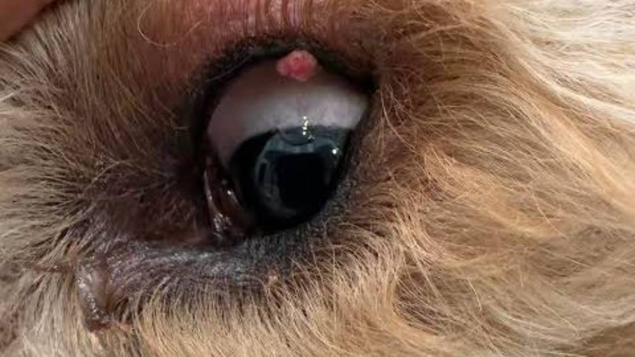
A meibomian gland adenoma is a benign tumor that grows from the oil glands along the edge of a dog's eyelid. It's one of the most common eyelid growths in dogs, usually appears in older dogs, and does not spread to other parts of the body (Gelatt, 2024; Hamor, 2024). In a study of 119 eyelid masses removed from dogs, meibomian adenoma was the second most common diagnosis, and 92.4% of all the masses turned out not to be cancer (Wang et al., 2019).
Even though it's benign, a meibomian gland adenoma can still cause trouble. As it grows, it can rub against the eye and irritate it, which is the most common reason to remove one (Gelatt, 2024; Hamor, 2024). A small number of eyelid lumps that look similar are malignant, so the lump should be removed and examined, or at least monitored by your veterinarian, rather than assumed harmless.
Key points
- Meibomian glands make the oily layer of the tear film. Their tumors are the most common eyelid tumors in older dogs (Hamor, 2024).
- Most eyelid masses in dogs are benign. Studies have found that 88% to over 90% are not cancer (Roberts et al., 1986, as cited in Wang et al., 2019; Wang et al., 2019).
- The malignant version, meibomian gland adenocarcinoma, can invade nearby tissue but isn't known to spread elsewhere in the body (Gelatt, 2024).
- Treatment is usually surgical removal, or removal of most of the mass followed by freezing the base (cryotherapy). Laser removal is another option (Bussieres et al., 2005; Hamor, 2024).
- After cryotherapy, adenomas were less likely to come back than other eyelid mass types in one study (Zibura et al., 2019).
- Some eyelid lumps in dogs are melanomas or mast cell tumors. A biopsy is the only way to know which kind your dog has (Gelatt, 2024; Hamor, 2024).
Detailed explanation
What meibomian glands are
Each eyelid has a row of small oil glands, the meibomian glands, with openings along the lid margin. The oil they release helps keep tears from evaporating. When cells in one of these glands grow abnormally, they can form a small bump right at the edge of the lid.
Pathologists use a few different names for these growths, and you may see any of them on a report. A meibomian adenoma is a benign tumor of the gland. A meibomian epithelioma is a closely related benign tumor; in the study of 119 masses, it was actually the most common diagnosis (34.5%), with adenoma close behind (29.4%), followed by meibomian hyperplasia, a non-cancerous overgrowth (18.5%) (Wang et al., 2019). A meibomian adenocarcinoma is the malignant form, and it was much less common, found in 6 of 119 masses (Wang et al., 2019).
How often eyelid lumps in dogs are cancer
The reassuring part is how often they aren't. In Wang et al.'s (2019) series of 119 eyelid masses from 118 dogs, 67.2% were benign tumors, 25.2% weren't tumors at all, and 7.6% were malignant. An older study of 200 dogs reported that 88% of eyelid tumors were benign (Roberts et al., 1986, as cited in Wang et al., 2019).
Size can be a clue. Malignant masses in the Wang study were significantly larger than benign ones (Wang et al., 2019). It's a clue only, though; a small lump can still be malignant, and a large one can be benign.
Even meibomian adenocarcinoma tends to behave locally. It can invade surrounding eyelid tissue and cause harm there, but it isn't known to spread to distant parts of the body (Gelatt, 2024; Hamor, 2024).
Which other eyelid lumps it can be confused with
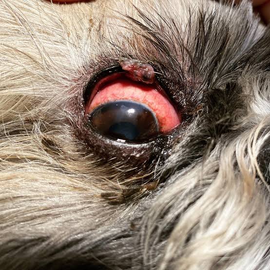
Several other growths show up on dogs' eyelids, including histiocytomas, papillomas, melanomas, and mast cell tumors (Gelatt, 2024; Hamor, 2024). Some of these matter more than others.
A histiocytoma is a benign growth that often goes away on its own. Eyelid melanomas are usually treated with wide removal or debulking with cryotherapy (Hamor, 2024). Mast cell tumors are always a form of cancer and range from easily cured to aggressive, so finding one on the eyelid changes the plan considerably.
You can't reliably tell these apart by looking. That's why your veterinarian will want tissue examined. Fine-needle aspirates, which work well for many skin lumps, are often non-diagnostic for eyelid masses in dogs (Geller, 2009), so the answer usually comes from a biopsy of the removed tissue. Our guide to reading a pathology report explains what you'll see on the results.
Which dogs get them
Eyelid tumors are mostly found in older dogs (Hamor, 2024; Roberts et al., 1986, as cited in Kaya et al., 2018). Roberts and colleagues also reported that Beagles, Siberian Huskies, and English Setters were more commonly affected, while mixed-breed dogs were less at risk (Roberts et al., 1986, as cited in Kaya et al., 2018).
Signs to watch for
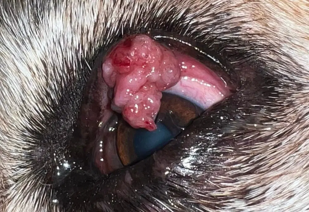
The lump itself is usually the first thing owners notice: a small bump on the edge of the upper or lower lid. Eyelid tumors are irritating to the dog and can be disfiguring (Gelatt, 2024). As the mass grows, it can rub on the surface of the eye. Signs that the eye is being irritated include squinting, extra tearing or discharge, redness, and pawing or rubbing at the face.
Local irritation is the main reason treatment becomes necessary (Hamor, 2024). If your dog has a lid lump and any of those signs, don't wait for the next routine visit.
Treatment options
Most eyelid tumors in dogs can be treated successfully with surgical excision, or with debulking and cryotherapy (Hamor, 2024). The right choice depends on the size and location of the mass, your dog's health, and whether general anesthesia is a concern.
Surgical excision. The mass is cut out along with a margin of eyelid, and the lid is stitched closed. Removing the whole mass gives the pathologist a complete sample and can confirm the margins. Our article on whether clear margins mean your dog is in the clear covers what that part of the report means.
Debulking with cryotherapy. The visible part of the mass is trimmed off and the base is frozen to destroy remaining cells. In a University of Minnesota study of 46 eyelid masses in 31 dogs treated this way, 15.2% recurred, at an average of about a year later. Recurrence depended on tumor type: adenomas were significantly less likely to recur, while mixed adenoma-papillomas were more likely to (Zibura et al., 2019). Some dogs can have this done under local anesthesia with little or no sedation. In a study of 38 eyelid masses in 30 dogs treated that way, 26.3% recurred, and no surgical complications were reported beyond minor hair loss, depigmentation, and mild inflammation (Han & Kim, 2022). That option may be worth asking about for older dogs or dogs with other health problems.
Carbon dioxide laser. In a study of 12 dogs, each with a meibomian gland adenoma, laser ablation under brief general anesthesia produced no recurrences at the six-month recheck. The authors concluded that it's an effective alternative to standard surgery (Bussieres et al., 2005). This study was small and short, and not every practice has the equipment.
Recurrence is possible with any method. A veterinary ophthalmologist writing on eyelid tumors suggests expecting it roughly 10% to 15% of the time (Geller, 2009). A lump that returns at the same spot usually needs another procedure, and new lumps can form at other glands.
Should every meibomian adenoma be removed?
Not necessarily right away. A very small, unchanging lump that isn't touching the eye may simply be monitored, and that decision is one to make with your veterinarian. Irritation of the eye is the clearest reason to act (Hamor, 2024). Size still matters, though. Once a mass involves more than about a third of the lid's length, removing it calls for reconstructive eyelid surgery rather than a simple excision (Geller, 2009). Measure the lump or take a dated photo every few weeks so you can show your veterinarian exactly how it's changing. Our 10-minute dog cancer check is an easy routine for tracking lumps anywhere on the body.
When to contact a veterinarian
Book an appointment for any new lump on or near your dog's eyelid, especially in an older dog, and for any eyelid lump that grows, changes color, bleeds, or ulcerates.
Call the same day if your dog is squinting, holding the eye closed, pawing at it, or has a cloudy patch or new discharge from the eye. These can mean the mass is damaging the surface of the eye, and that can become painful quickly.
Questions to ask your veterinarian
- Do you think this is a meibomian gland tumor, or could it be something else?
- Is it touching or irritating the eye right now?
- Should we remove it now or monitor it, and how fast would it need to change before we act?
- Would you recommend excision, debulking with cryotherapy, or laser removal for this mass, and why?
- Could this be done under local anesthesia or light sedation instead of general anesthesia?
- Will the removed tissue be sent for a biopsy?
- If it's an adenoma, what are the chances it comes back?
- Should my dog see a veterinary ophthalmologist for this?
Sources
Bussieres, M., Krohne, S. G., Stiles, J., & Townsend, W. M. (2005). The use of carbon dioxide laser for the ablation of meibomian gland adenomas in dogs. Journal of the American Animal Hospital Association, 41(4), 227-234.
Gelatt, K. N. (2024). Cancers and tumors of the eye in dogs. Merck Veterinary Manual, Dog Owners Version. Retrieved November 5, 2026, from https://www.merckvetmanual.com/dog-owners/eye-disorders-of-dogs/cancers-and-tumors-of-the-eye-in-dogs
Geller, A. (2009, April 1). Ocular neoplasia and treatment [Conference proceedings]. dvm360. https://www.dvm360.com/view/ocular-neoplasia-and-treatment-proceedings
Hamor, R. E. (2024). Ocular neoplasia in dogs. Merck Veterinary Manual, Professional Version. Retrieved November 5, 2026, from https://www.merckvetmanual.com/eye-diseases-and-disorders/neoplasia-of-the-eye-and-associated-structures/ocular-neoplasia-in-dogs
Han, M.-G., & Kim, J.-Y. (2022). Application of local anaesthesia and cryosurgery for eyelid masses in dogs. Veterinary Medicine and Science, 8, 476-482.
Kaya, D. A., Güzel, Ö., Sevim, G., Demir, A., Erdikmen, D. O., Öztürk, G. Y., & Bamaç, Ö. E. (2018). Eyelid tumors in dogs: 47 cases retrospective study (2006-2017). Kocatepe Veterinary Journal, 11(3), 286-291.
Wang, S. L., Dawson, C., Wei, L. N., & Lin, C. T. (2019). The investigation of histopathology and locations of excised eyelid masses in dogs. Veterinary Record Open, 6(1), Article e000344.
Zibura, A. E., Henriksen, M. de L., Rendahl, A., Lim, C. C., & Reilly, C. (2019). Retrospective evaluation of canine palpebral masses treated with debulking and cryotherapy: 46 cases. Veterinary Ophthalmology, 22(3), 256-264.
Reviewed by: Amber L. Drake, PhD
Dr. Amber L. Drake is a board-certified holistic health practitioner, canine clinical herbalist, educator, and founder of the Drake Dog Cancer Foundation and Drake Dog Academy. She is dedicated to helping pet parents better understand canine cancer, treatment options, nutrition, quality of life, and supportive care through compassionate, evidence-informed education. Her work combines professional training, practical resources, and firsthand insight from supporting thousands of dog families through the challenges of a cancer diagnosis.
Stay connected with news and updates!
Join our mailing list to receive the latest news and updates from our team.
Don't worry, your information will not be shared.
We hate SPAM. We will never sell your information, for any reason.