Are All Mast Cell Tumors Cancerous in Dogs?
Nov 05, 2026
Yes. Every mast cell tumor in a dog is a form of cancer. Veterinary specialists don't classify any canine mast cell tumor as benign, and an international consensus group describes mast cell neoplasms as one of the most frequently diagnosed malignancies in dogs (Willmann et al., 2021). Penn Vet's oncology service puts it simply: a mast cell tumor "represents a cancer of a type of blood cell" (Penn Vet Ryan Hospital, 2017).
That word "cancer" covers a very wide range, though. Many low-grade mast cell tumors are cured by surgery alone, while high-grade tumors can spread and shorten life considerably. In a study that helped set the grading system pathologists use today, dogs with low-grade tumors had a median survival of more than two years, compared with less than four months for high-grade tumors (Kiupel et al., 2011). The grade, location, and stage of your dog's tumor matter far more than the label.
Key points
- All mast cell tumors in dogs are cancer. Some behave so gently that specialists describe their behavior as benign, but the tumor itself is never classified that way (Willmann et al., 2021).
- They're common, making up roughly 20% of canine skin tumors (Harshbarger & Meinkoth, 2023).
- A biopsy grade is the single most important piece of information. Pathologists sort tumors into low grade and high grade (Kiupel et al., 2011).
- Low grade doesn't guarantee good behavior. Around 15% of low-grade tumors act more aggressively than expected (Willmann et al., 2021).
- Tumors that sit under the skin, rather than in it, usually have a favorable outlook. In one study of 306 dogs, only 4% developed metastasis (Thompson et al., 2011).
- Mast cell tumors can release substances that cause swelling, redness, and stomach ulcers, which is why they sometimes seem to change size from day to day (Penn Vet Ryan Hospital, 2017).
Detailed explanation
Why "cancer" and "benign behavior" can both be true
Mast cells are immune cells that normally help with allergic and inflammatory responses. A mast cell tumor forms when these cells multiply out of control (Penn Vet Ryan Hospital, 2017; Willmann et al., 2021). Because they're tumors of a blood cell type that grow without the body's normal limits, they're considered malignant by definition.
Malignant describes what kind of tumor it is. It doesn't tell you how a particular tumor will act. The consensus group that proposed diagnostic criteria for canine mast cell neoplasms recommends against full staging after removal of most low-grade skin tumors "due to the benign biological behavior of the disease" (Willmann et al., 2021). That's the source of a lot of confusion. Your veterinarian may tell you the tumor is cancer and, in the same conversation, say it's unlikely to cause your dog any more trouble once it's removed. Both statements can be accurate.
The same paper is careful not to stretch that reassurance too far. It notes that around 15% of tumors graded low by the current system may behave more aggressively, and it never calls any canine mast cell neoplasm benign outright (Willmann et al., 2021).
What grade tells you
Grade describes how abnormal the cells look under the microscope and how fast they appear to be dividing. For years, pathologists used a three-tier system, but agreement between pathologists on the middle grade was poor. When 28 pathologists graded the same 95 tumors, they agreed on the older grades 1 and 2 less than 64% of the time (Kiupel et al., 2011).
The two-tier system that came out of that work is now widely used. A tumor is called high grade if it has any one of these features: at least 7 dividing cells in 10 microscope fields, at least 3 cells with three or more nuclei, at least 3 bizarre nuclei, or marked variation in nuclear size in at least 10% of cells. Otherwise it's low grade (Kiupel et al., 2011).
The difference in outcome is large. High-grade tumors were significantly linked to a shorter time before the cancer spread or new tumors appeared, and median survival was under 4 months for high grade versus over 2 years for low grade (Kiupel et al., 2011). Our article on mast cell tumor life expectancy goes further into what shapes survival.
Your biopsy report may list both grading systems. Ask your veterinarian to walk you through each line; our guide to reading a pathology report can help you prepare.
Where the tumor sits matters too
Some mast cell tumors grow in the skin itself (cutaneous). Others grow in the fatty tissue underneath (subcutaneous). The subcutaneous form usually behaves better. In a study of 306 dogs with subcutaneous mast cell tumors, 8% had the tumor come back at the surgery site, 4% developed metastasis, and 9% died of mast cell disease. The authors concluded that "the majority of subcutaneous MCTs have a favorable prognosis" (Thompson et al., 2011).
That's a tendency rather than a promise. Subcutaneous tumors can still recur or spread, so location alone can't predict how a tumor will behave (Willmann et al., 2021). Mast cell disease that has spread to the lymph nodes and beyond, or that arises in internal organs such as the spleen, liver, or intestinal tract, generally carries a poor prognosis (Penn Vet Ryan Hospital, 2017).
Breed can shift the odds
In an analysis of 9,375 confirmed skin mast cell tumors from U.S. dogs, the proportion of high-grade tumors differed significantly between breeds. Pugs had a higher risk of low- and intermediate-grade tumors but not of high-grade ones, meaning their tumors tended to be the less aggressive kind. Staffordshire Bull Terriers, American Staffordshire Terriers, and Boxers, on the other hand, had a higher relative risk of high-grade tumors (Mochizuki et al., 2017). The authors suggest genetics play a part in which dogs develop high-grade disease.
A breed tendency is only a starting point. A Pug can still have a high-grade tumor, and the biopsy is what settles it.
Why a mast cell tumor can't be judged by how it looks
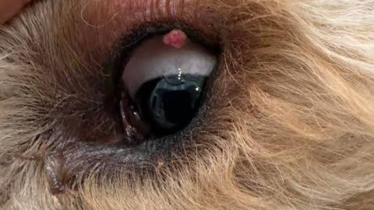
Mast cell tumors look like almost anything. They may be a raised lump in or under the skin, and they can be red, ulcerated, or swollen. Some sit nearly unchanged for months, while others appear suddenly and grow quickly (Penn Vet Ryan Hospital, 2017). A soft lump that seems to be "just a fatty lump" can turn out to be a mast cell tumor, which is why vets aspirate lumps rather than guess. If you're trying to tell lumps apart at home, our guide on how to identify mast cell tumors and our overview of lumps that aren't cancer are useful, but neither replaces a needle sample.
Many owners notice the lump growing and shrinking. That happens because mast cells contain granules that can be released, causing local swelling and redness, sometimes on their own and sometimes after the tumor is handled or bumped. Released granule contents can also cause stomach ulcers and bleeding and, in severe cases, a dangerous drop in blood pressure (Penn Vet Ryan Hospital, 2017). It's best not to squeeze or repeatedly press on a lump you suspect.
How the diagnosis and grade are found
Most mast cell tumors are first identified with a fine-needle aspirate, a quick test in which cells are drawn out with a small needle (Harshbarger & Meinkoth, 2023; Penn Vet Ryan Hospital, 2017). Cytology can also suggest a grade. In one study, a cytology grading scheme identified high-grade tumors with 88% sensitivity and 94% specificity compared with biopsy (Camus et al., 2016).
Biopsy is still the gold standard for grading. A tissue sample also lets the pathologist see whether the tumor is in or under the skin and, after surgery, whether the margins are clear, which cytology can't do (Harshbarger & Meinkoth, 2023). If you're worried about the procedure itself, here's whether a fine-needle aspirate hurts.
Depending on what the aspirate shows, your veterinarian may also recommend staging tests, such as lymph node aspirates, abdominal ultrasound, and bloodwork, to check whether the cancer has spread (Penn Vet Ryan Hospital, 2017).
What this means for treatment
Surgery is the treatment of choice for skin mast cell tumors, and complete removal of a low-grade tumor may be curative. When margins aren't clean, a second surgery or radiation therapy may be recommended; radiation provides high rates of long-term control at the original site. High-grade tumors and those that have spread usually need additional treatment, such as chemotherapy, steroids, or a targeted drug like Palladia, alongside local treatment (Penn Vet Ryan Hospital, 2017). Clean margins are good news, but they aren't the whole story; our article on whether clear margins mean your dog is in the clear explains why follow-up still matters.
When to contact a veterinarian
Make an appointment for any new lump, any lump that changes size, color, or texture, and any lump that swells, reddens, or ulcerates. A lump that grows and shrinks is worth checking soon, since that pattern is typical of mast cell tumors (Penn Vet Ryan Hospital, 2017).
Call your veterinarian the same day if your dog has a known or suspected mast cell tumor and starts vomiting, has black or tarry stool, or stops eating, because granule release can cause stomach ulcers and bleeding (Penn Vet Ryan Hospital, 2017). Go to an emergency hospital right away if your dog collapses, has very pale gums, or develops sudden widespread swelling, hives, or weakness, especially after a lump has been handled or removed.
Questions to ask your veterinarian
- Was this diagnosed by aspirate or biopsy, and has it been graded?
- What grade is it under the two-tier system, and what did the older three-tier system say if both were reported?
- Is the tumor in the skin or under the skin?
- Were the surgical margins clear, and how wide were they?
- Do you recommend staging tests before or after surgery?
- Should a lymph node near the tumor be checked or removed?
- Is my dog at risk for stomach ulcers from this tumor, and are there signs I should watch for?
- Would it help to see a veterinary oncologist before deciding on treatment?
- How often should we recheck for new lumps, given that dogs can develop more than one?
Sources
Camus, M. S., Priest, H. L., Koehler, J. W., Driskell, E. A., Rakich, P. M., Ilha, M. R., & Krimer, P. M. (2016). Cytologic criteria for mast cell tumor grading in dogs with evaluation of clinical outcome. Veterinary Pathology, 53(6), 1117-1123.
Harshbarger, A. A., & Meinkoth, J. H. (2023, February 13). Cytologic grading of mast cell tumors in small animals. Today's Veterinary Practice. https://todaysveterinarypractice.com/clinical-pathology/cytologic-grading-of-mast-cell-tumors-in-small-animals/
Kiupel, M., Webster, J. D., Bailey, K. L., Best, S., DeLay, J., Detrisac, C. J., Fitzgerald, S. D., Gamble, D., Ginn, P. E., Goldschmidt, M. H., Hendrick, M. J., Howerth, E. W., Janovitz, E. B., Langohr, I., Lenz, S. D., Lipscomb, T. P., Miller, M. A., Misdorp, W., Moroff, S., . . . Miller, R. (2011). Proposal of a 2-tier histologic grading system for canine cutaneous mast cell tumors to more accurately predict biological behavior. Veterinary Pathology, 48(1), 147-155.
Mochizuki, H., Motsinger-Reif, A., Bettini, C., Moroff, S., & Breen, M. (2017). Association of breed and histopathological grade in canine mast cell tumours. Veterinary and Comparative Oncology, 15(3), 829-839.
Penn Vet Ryan Hospital. (2017). Mast cell tumors in dogs [Client information sheet]. Comprehensive Cancer Care Service, University of Pennsylvania School of Veterinary Medicine. https://www.vet.upenn.edu/wp-content/uploads/2025/05/mast-cell-tumors-in-dogs.pdf
Thompson, J. J., Pearl, D. L., Yager, J. A., Best, S. J., Coomber, B. L., & Foster, R. A. (2011). Canine subcutaneous mast cell tumor: Characterization and prognostic indices. Veterinary Pathology, 48(1), 156-168.
Willmann, M., Yuzbasiyan-Gurkan, V., Marconato, L., Dacasto, M., Hadzijusufovic, E., Hermine, O., Sadovnik, I., Gamperl, S., Schneeweiss-Gleixner, M., Gleixner, K. V., Böhm, T., Peter, B., Eisenwort, G., Moriggl, R., Li, Z., Jawhar, M., Sotlar, K., Jensen-Jarolim, E., Sexl, V., . . . Valent, P. (2021). Proposed diagnostic criteria and classification of canine mast cell neoplasms: A consensus proposal. Frontiers in Veterinary Science, 8, Article 755258.
Reviewed by: Amber L. Drake, PhD
Dr. Amber L. Drake is a board-certified holistic health practitioner, canine clinical herbalist, educator, and founder of the Drake Dog Cancer Foundation and Drake Dog Academy. She is dedicated to helping pet parents better understand canine cancer, treatment options, nutrition, quality of life, and supportive care through compassionate, evidence-informed education. Her work combines professional training, practical resources, and firsthand insight from supporting thousands of dog families through the challenges of a cancer diagnosis.
Stay connected with news and updates!
Join our mailing list to receive the latest news and updates from our team.
Don't worry, your information will not be shared.
We hate SPAM. We will never sell your information, for any reason.