Sebaceous Cysts on Dogs: What They Are and When to Worry
Nov 05, 2026
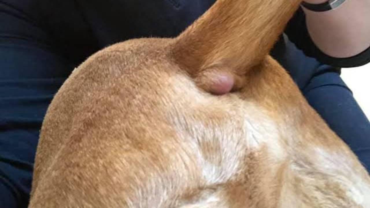
Most of what people call sebaceous cysts on dogs are follicular cysts: small, benign sacs that form in or under the skin and fill up with keratin, the same protein that makes up hair and the outer skin layer. True cysts of the sebaceous (oil) glands are extremely rare in dogs (Cortadellas, n.d.). Follicular cysts usually feel like smooth, round, firm or squishy lumps, typically between about half a centimeter and 5 centimeters across, and they're most often found on the head, neck, trunk, and front legs (Cortadellas, n.d.).
These cysts aren't cancer, and surgical removal cures them completely (Huber & Beck, 2017). The catch is that they can look and feel like other lumps, including tumors, so a veterinarian should check any new lump with a needle sample before you assume it's "just a cyst" (Huber & Beck, 2017). And don't squeeze one. Pressing out the contents can push keratin into the surrounding skin and set off a painful inflammatory reaction (Cortadellas, n.d.; H.-J. Kim et al., 2021).
Key points
- "Sebaceous cyst" is a common name, not an accurate one. In dogs, these lumps are almost always follicular cysts filled with keratin, not oil (Cortadellas, n.d.; H.-J. Kim et al., 2021).
- Follicular cysts are benign. Removing them surgically is curative (Huber & Beck, 2017).
- They can mimic both benign and malignant skin tumors, so diagnosis needs cytology or a biopsy (Huber & Beck, 2017).
- Mast cell tumors are the most common skin tumor in dogs in a large Swiss registry, which is one reason lumps shouldn't be judged by touch alone (Graf et al., 2018).
- Squeezing a cyst or having it rupture can trigger swelling, pain, and sometimes infection (Cortadellas, n.d.; Huber & Beck, 2017).
- Not every cyst needs surgery. Monitoring can be reasonable once the diagnosis is confirmed, especially for dogs with many cysts (Cortadellas, n.d.).
Detailed explanation
Why "sebaceous cyst" is usually the wrong name
Sebaceous glands sit beside hair follicles and make sebum, the skin's natural oil. It's easy to assume a cyst full of thick, cheesy material must come from those glands. In dogs, it almost never does. What owners and even many veterinarians call a sebaceous cyst is usually a follicular cyst, most often the infundibular type, which forms from the upper part of the hair follicle (Cortadellas, n.d.).
The difference shows up in what's inside. Follicular cysts are packed with keratin, arranged in layers in the most common type (Cortadellas, n.d.). H.-J. Kim and colleagues (2021) likewise describe the central contents of these cysts as keratin rather than sebum. Pathologists classify follicular cysts further by which part of the follicle they come from, using names like infundibular, isthmic, matrical, or hybrid cysts (Cortadellas, n.d.; Huber & Beck, 2017). If your dog's biopsy report uses one of these names, it's describing the same kind of lump you may have heard called a sebaceous cyst.
Genuine sebaceous gland cysts do exist but are extremely rare in dogs, and they tend to be firm, single nodules smaller than 1 centimeter (Cortadellas, n.d.).
How they form and what they look like
A follicular cyst develops when the opening of a hair follicle gets blocked and the follicle keeps producing keratin with nowhere to go. Chronic friction can contribute (H.-J. Kim et al., 2021). The cyst grows slowly as keratin builds up, stretching and thinning its wall over time (Huber & Beck, 2017).
Most are single, firm lumps in the skin, occasionally deeper under it (H.-J. Kim et al., 2021). Some dogs develop several at once, particularly along the back or over pressure points such as elbows, and multiple cysts have been linked to pressure or trauma in larger breeds (Cortadellas, n.d.; H.-J. Kim et al., 2021). A cyst may stay the same size for a long time or slowly enlarge (H.-J. Kim et al., 2021).
They can show up in young dogs too. In a study of more than 2,500 skin masses removed from dogs under a year old, follicular cysts made up 1.7% of diagnoses, behind histiocytomas, papillomas, and dermoid cysts (D. Kim et al., 2022).
Why squeezing a cyst is a bad idea
It's tempting to pop a cyst like a pimple, especially when it already has a small opening and some material is coming out. The problem is that pressure can rupture the cyst wall below the surface and push its contents into the surrounding skin. The body reacts to that loose keratin as if it were a foreign object (Cortadellas, n.d.).
That reaction can be dramatic. Huber and Beck (2017) describe owners finding a swelling as large as a fist within 24 to 48 hours, at a spot where only a small nodule had been before. In two dogs reported by H.-J. Kim and colleagues (2021), ruptured cysts became painful, red, enlarged, and in one case crusted and bleeding, and the inflammation needed medical treatment before the cysts could be dealt with. A ruptured cyst can also become infected (Cortadellas, n.d.).
If a cyst has opened on its own, keep the area clean, keep your dog from licking or scratching it, and have your veterinarian look at it.
Why a lump shouldn't be assumed to be a cyst
Follicular cysts can resemble both benign and malignant skin tumors, which is why cytology and histopathology are needed to tell them apart (Huber & Beck, 2017). This matters because skin tumors are common in dogs. In a Swiss registry of 11,740 canine skin tumors, the most common type was the mast cell tumor (16.35%), followed by lipomas, hair follicle tumors, histiocytomas, and soft tissue sarcomas (Graf et al., 2018).
Mast cell tumors are a particular concern because every one of them is a form of cancer, even though many are cured by surgery. Our guide on how to identify mast cell tumors explains why touch and appearance aren't enough.
The test that usually settles it is a fine-needle aspirate, in which a small needle draws cells or contents out of the lump for examination under a microscope. A follicular cyst typically yields keratin and debris; a tumor yields its own kind of cells. When the aspirate isn't conclusive, a biopsy gives the definitive answer. If the procedure worries you, here's whether a fine-needle aspirate hurts.
Cysts versus sebaceous gland growths
Sebaceous glands do give rise to a different, common group of lumps: sebaceous hyperplasia, benign sebaceous tumors, and, less often, malignant ones. Many are benign and unlikely to spread, but they can still be locally aggressive and often rupture into sores that don't heal, which usually means surgical removal; cytology helps guide the next step, and a biopsy is needed for a definitive diagnosis (Merck Veterinary Manual, n.d.). If your veterinarian mentions a "sebaceous adenoma," that's a growth of the gland itself, not a cyst. A related growth on the eyelid margin is covered in our article on meibomian gland adenomas.
Treatment: removal or monitoring
Once a lump is confirmed to be a follicular cyst, there are two reasonable paths. Surgical removal of the whole cyst, including its wall, cures it (Huber & Beck, 2017), and it's the recommended treatment for cutaneous cysts in dogs (H.-J. Kim et al., 2021). Removing a cyst before it ruptures is usually simpler, because rupture and the inflammation that follows make surgery more difficult (H.-J. Kim et al., 2021).
Monitoring is also a valid choice in some cases, particularly for dogs with many cysts scattered over the body, where extensive surgery for benign lumps may not be justified (Cortadellas, n.d.). The decision should be made with your veterinarian based on the size, location, number, and behavior of the cysts (Cortadellas, n.d.).
If you're monitoring, track each lump. Note its location, measure it, and take a dated photo every month. Our 10-minute dog cancer check gives you a simple routine, and our overview of lumps other than cancer covers the other common harmless masses.
When to contact a veterinarian
Book an appointment for:
- Any new lump that hasn't been checked with a needle sample
- A known cyst that grows, changes texture, or starts to look different
- More than one new lump appearing over a short time
Call the same day if:
- A lump suddenly swells, turns red, or becomes painful
- A cyst has opened and is draining pus, bleeding, or smells bad
- Your dog won't stop licking, chewing, or scratching at a lump
Questions to ask your veterinarian
- Can we do a fine-needle aspirate to confirm this is a cyst?
- If it's a follicular cyst, is there any reason to remove it now, or can we monitor it?
- Is it in a spot where it's likely to be bumped, rubbed, or rupture?
- If it ruptures, what should I do at home before I can bring my dog in?
- Should the removed tissue be sent for a biopsy?
- My dog has several lumps. Do all of them need to be sampled, or only certain ones?
- How should I track these lumps between visits?
Sources
Cortadellas, O. (n.d.). Sebaceous cysts in dogs. Are they really sebaceous? Vets & Clinics. Retrieved November 5, 2026, from https://vetsandclinics.com/en/library/sebaceous-cysts-in-dogs-are-they-really-sebaceous
Graf, R., Pospischil, A., Guscetti, F., Meier, D., Welle, M. M., & Dettwiler, M. A. (2018). Cutaneous tumors in Swiss dogs: Retrospective data from the Swiss Canine Cancer Registry, 2008-2013. Veterinary Pathology, 55(6), 809-820. https://doi.org/10.1177/0300985818789466
Huber, D., & Beck, A. (2017). Canine cutaneous cysts of follicular epithelial origin. Hrvatski veterinarski vjesnik, 25(1/2), 72-89. https://hrcak.srce.hr/en/clanak/336262
Kim, D., Dobromylskyj, M. J., O'Neill, D., & Smith, K. C. (2022). Skin masses in dogs under one year of age. Journal of Small Animal Practice, 63(1), 10-15.
Kim, H.-J., Cho, K.-O., & Baek, Y.-B. (2021). Foreign body reaction to ruptured follicular cysts in dogs. Veterinary Medicine and Science, 7, 1509-1513.
Merck Veterinary Manual. (n.d.). Irish terrier with mass on head [Case study]. Retrieved November 5, 2026, from https://www.merckvetmanual.com/iframe/manual/case-studies/case006-vet/about.html
Reviewed by: Amber L. Drake, PhD
Dr. Amber L. Drake is a board-certified holistic health practitioner, canine clinical herbalist, educator, and founder of the Drake Dog Cancer Foundation and Drake Dog Academy. She is dedicated to helping pet parents better understand canine cancer, treatment options, nutrition, quality of life, and supportive care through compassionate, evidence-informed education. Her work combines professional training, practical resources, and firsthand insight from supporting thousands of dog families through the challenges of a cancer diagnosis.
Stay connected with news and updates!
Join our mailing list to receive the latest news and updates from our team.
Don't worry, your information will not be shared.
We hate SPAM. We will never sell your information, for any reason.